文献:PD-L1-targeted microbubbles loaded with docetaxel produce a synergistic effect for the treatment of lung cancer under ultrasound irradiation
文献链接:https://pubmed.ncbi.nlm.nih.gov/31942578/
作者:Tiankuan Li, Zhongqian Hu, Chao Wang, Jian Yang, Chuhui Zeng , Rui Fan, Jinhe Guo
相关产品:DSPE-PEG-FITC
原文摘要:Immunotherapy is gradually becoming as important as traditional therapy in the treatment of cancer, but adverse drug reactions limit patient benefits from PD1/PD-L1 checkpoint inhibitor drugs in the treatment of non-small cell lung cancer (NSCLC). As a chemotherapeutic drug for NSCLC, docetaxel (DTX) can synergize with PD1/PD-L1 checkpoint inhibitors but increase haematoxicity and neurotoxicity. Herein, anti-PDL1 monoclonal antibody (mAb)-conjugated and docetaxel-loaded multifunctional lipid-shelled microbubbles (PDMs), which were designed with biological safe phospholipid to produce synergistic antitumour effects, reduced the incidence of side effects and promoted therapeutic effects under ultrasound (US) irradiation. The PDMs were prepared by the acoustic-vibration method and then conjugated with an anti-PDL1 mAb. The material features of the microbubbles, cytotoxic effects, cellular apoptosis and cell cycle inhibition were studied. A subcutaneous tumour model was established to test the drug concentration-dependent and antitumour effects of the PDMs combined with US irradiation, and an orthotopic lung tumour model simultaneously verified the antitumour effect of this synergistic treatment. The PDMs achieved higher cellular uptake than free DTX, especially when combined with US irradiation. The PDMs combined with US irradiation also induced an increased rate of cellular apoptosis and an elevated G2-M arrest rate in cancer cells, which was positively correlated with PDL1 expression. An in vivo study showed that synergistic treatment had relatively strong effects on tumour growth inhibition, increased survival time and decreased adverse effect rates. Our study possibly provides a well-controlled design for immunotherapyand chemotherapy and has promising potential as a clinical application for NSCLC treatment.
超声照射可以暂时性地增加细胞膜的通透性,这被称为声孔效应。这种效应允DSPE-PEG-FITC包裹的DTX更容易地进入细胞内,从而提高药物的摄取效率。DSPE-PEG-FITC形成的脂质体具有良好的稳定性,能够在血液循环中长时间存在,减少药物的非特异性分布。FITC荧光标记使得DSPE-PEG-FITC包裹的DTX可以通过荧光显微镜等设备进行实时观察和追踪,有助于研究药物在细胞内的分布和动态行为。DSPE-PEG-FITC脂质体可以在细胞内释放DTX,通过改变脂质体的结构和交联程度来调节药物的释放速率,实现药物的可控释放。

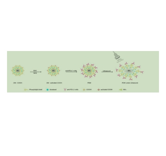
图为:DTX和抗pd-l1单抗共负载微气泡的制备示意图。
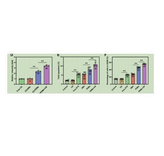
DSPE-PEG-FITC对于荧光成像和流式细胞术在不同配方中DTX的细胞摄取:
一、荧光成像测定DTX的细胞摄取
细胞培养:将细胞培养在适宜的培养基中,确保细胞处于对数生长期,状态良好。
荧光标记DTX:将DTX与荧光染料(如FITC)结合,制备成荧光标记的DTX。
细胞处理:将不同配方的荧光标记DTX加入细胞培养液中,与细胞共孵育一定时间。
洗涤细胞:去除未进入细胞的药物,用PBS等缓冲液洗涤细胞。
荧光成像:使用荧光显微镜或共聚焦显微镜观察细胞内的荧光信号,记录荧光强度和分布。
二、流式细胞术测定DTX的细胞摄取
细胞处理:将细胞培养在适宜的培养基中,然后收集细胞,并用不同配方的DTX进行处理。
荧光标记:将DTX与荧光染料结合,或者直接使用荧光标记的抗体来标记细胞表面的DTX受体。
洗涤细胞:去除未结合的荧光染料或抗体,用PBS等缓冲液洗涤细胞。
流式细胞仪检测:将处理后的细胞悬液注入流式细胞仪中,使用激光激发荧光染料,检测细胞的荧光强度。
数据分析:根据流式细胞仪的数据,分析不同配方中DTX的细胞摄取情况。

图为:经超声照射、游离DTX、DMs、PDMs或PDMs经超声照射处理后的LLC细胞的细胞周期抑制研究。
结论:DSPE-PEG-FITC在超声照射下摄取DTX的机制是通过超声增强的细胞膜渗透性提高药物摄取,结合脂质体的稳定性和靶向递送能力,以及荧光标记便于实时追踪和药物的可控释放。这些特性共同促进了DSPE-PEG-FITC在药物递送系统中的应用,是在提高药物Therapeutic effect 和减少副作用方面显示出潜力。

 2024-12-18 作者:lkr 来源:

