原文献:
不同游离脂肪酸通过PI3K/AKT途径对INS-1β细胞增殖与Apoptosis 的影响
文献链接:
https://xueshu.baidu.com/usercenter/paper/show?paperid=f1675505fcec2a4026863f152686c64c&site=xueshu_se&hitarticle=1
作者:
戴盈,崔巍,徐利,施秉银,赵廷启
原文摘要:Objective To investigate the efiect of'saturated fatty acid palmitate (PA)and unsaturated fatty acid Oleate(OA) on proliferation and apoptosis of lNS-1 β-cell., Methods Rat INS-1 β-cells were maintained in RPMI 1640,and then treated with different concentration of PA and OA. Cell proliferation was assessed by M'TT colorimetric assay. Inverted microscope was used to observe the morphological changes of FFAs-treated cells. The flow cytometrwas used to detect the FFAs-induced apoptosis oflNS-1 B cells by using double labeled Annexin-V/FITC/PI. Results
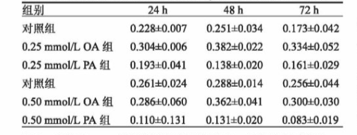
Compared with the control group, both concentrations ofOA promoted the growth of INs-1β cells, and 0.25 mmol/LOA could significantly promoted cell growth (P < 0.05). Both concentrations ofPA inhibited the growth ofINS-1βcells. Among them, 0.50 mmol/L, PA had a significant inhibitory effect (P< 0.01). Z-VAD-fmk could inhibit the in-hibitory effect of PA on the growth of INS-1β cells. LY294002 could promote the inhibitory effect of PA on the growthof INS-1β cells and inhibit the growth of INS-1β cells. OA could prevent apoptosis of INs-1p cells caused by highglucose environment, while PA c could significantly increase the proportion of INS-1β cell apoptosis. ConclusionINS-1 β cells exposed to PA under serum-free conditions showed marked apoptosis and little proliferation, howeverOA can protect IiNS-1 β cells. The apoptosis of INS-1 β cells exposed to high concentration and chronic PA was Caspase-dependent. Pl3K-Akt signal pathway plays an important role in lipoapoptosis of β cells by stimulating cell pro.liferation and inhibiting apoptosis.
FFA的升高可能与胰岛B细胞功能障碍相关,FFA 包括饱和脂肪酸棕榈酸(PA)和不饱和脂肪酸油酸(OA),不同 FFA对胰岛B细胞的生存与Apoptosis 的影响不同,该文献实验以大鼠 INS-1胰岛B细胞(以下简称INS-1细胞)为实验对象,观察不同浓度饱和脂肪酸(棕榈酸,PA)、单不饱和脂肪酸(油酸,OA)和 FFA加用广谱 Caspase 抑制剂和 PI3K 抑制剂对体外培养下的INS-1细胞的增殖的影响,采用MTT法检测细胞增殖,Annexin-V/FITC/PI双标记流式细胞术检测细胞。实验步骤如下:

图:不同浓度的油酸和棕榈酸对INS-1β细胞增殖(吸光度A)的影响
INS-1细胞用含10%胎牛血清的RPMI1640培养基在 37 ℃/5%CO2的细胞孵箱中培养,待到细胞 85%左右贴壁,弃去原有培养基,用无血清的含 0.1%BSA和 0.5mmol/葡萄糖的RPMI1640培养基对细胞进行24h同步化培养。然后用不同浓度的 PA 和 OA 进行干预。
用NaOH溶液在水浴中溶解OA 和 PA,振荡混匀过滤,配成OA/PA 储存液。在水浴中用去离子水配BSA 溶液,过滤。然后将上述的FFA 溶液和 BSA 溶液按体积比混合配成FFA/BSA复合液。
将INS-1细胞种于96孔培养板,待细胞贴壁,细胞同步化,弃去原有培养基,加入含不同浓度FFA的培养基干预。将INS-1细胞种于6孔培养板,待细胞贴壁,同步化培养。实验组分别用不同浓度的无血清高糖RPMI1640培养基培养,对照组用含BSA的无血清高糖RPMI1640培养基培养。在6、16、24h3个时间点,用倒置显微镜观察细胞并拍摄记录细胞形态,然后分别消化细胞,离心收集细胞,每个样本取细胞悬液,加入Annexin-V/FITC和碘化丙啶溶液,混匀后室温避光孵育,在每个样本中加入结合缓冲液,流式细胞仪测量并分析。

图:不同浓度 OA 和 PA 作用下对 INS-1β细胞Apoptosis 的影响
结论: PA 抑制 INS-1β细胞生长并促进其Apoptosis ,而 OA 对 INS-1细胞则具有保护作用;PA引起的细胞Apoptosis 有依赖 Caspase介导的Apoptosis 信号通路,而PI3K/AKt信号通路在胰岛B细胞脂性Apoptosis 中有维持应激细胞的生存的作用。

 2024-12-18 作者:ZJ 来源:https://xueshu.baidu.com/usercenter/paper/show?paperid=f1675505fcec2a4026863f152686c64c&site=xueshu_se&hitarticle=1

