
 2024-12-17 作者:lkr 来源:
文献:
A dual-signal amplification strategy for kanamycin based on ordered mesoporous carbon-chitosan/gold nanoparticles-streptavidin and ferrocene labelled DNA
文献链接:
https://www.sciencedirect.com/science/article/abs/pii/S0003267018307025
作者:
Falan Li , Xiangyou Wang , Xia Sun , Yemin Guo , Wenping Zhao
相关产品:
原文摘要:An ultrasensitive electrochemical aptasensor for kanamycin (KAN) detection was constructed with a dual-signal amplification strategy. The aptasensor achieved greatly amplified sensitivity due to the excellent electrical conductivity of the ordered mesoporous carbon-chitosan (OMC-CS)/gold nanoparticles-streptavidin (AuNPs-SA) and DNA2 labelled with ferrocene (Fc-DNA2). The AuNPs-SA was used to immobilize the DNA strand (biotin labelled) with the biotin-streptavidin system. The DNA2 strand containing the KAN aptamer was labelled with ferrocene to increase the current signal on the electrode surface when bound to KAN. Some factors that affect the performance of the aptasensor were optimized, and the proposed aptasensor provided a wide linear range from 1×10⁻¹⁰ M to 4×10⁻⁶ M, with a detection limit as low as 35.69 pM for KAN under the optimized conditions. This aptasensor had satisfactory electrochemical performance with good stability, sensitivity and reproducibility. Additionally, it also displayed a good specificity for KAN without interference from competitive analogues. Furthermore, the constructed aptasensor was successfully used to detect KAN in a real milk sample. The proposed method for KAN detection has great potential for the detection of other antibiotics.
采用双信号放大策略构建的卡那霉素(KAN)检测电化学适应传感器。由于有序介孔碳壳聚糖(OMC-CS)/金纳米颗粒链霉亲和素(AuNPs-SA)和标记二茂铁(Fc-DNA2)的DNA的优良电导率,该传感器放大灵敏度。AuNPs-SA用生物素-链霉亲和素系统固定DNA链(生物素标记)。将含有KAN适配体的条DNA链用二茂铁标记,以在与KAN结合时增加电极表面的电流信号。

图为:适应传感器组装程序的示意图
AuNPs-SA在适体传感器构建中的具体制备:
用氧化铝粉抛光,在乙醇、硝酸和双蒸馏水中进行超声洗涤。电极循环扫描,并放置在硫酸溶液中进行激活。然后,将CS-OMC悬浮液滴加到电极表面。电极干燥后,在其表面加入AuNPs-SA溶液。随后,将干燥的电极在 Bio-DNA1中孵育。然后,将电极浸泡在的牛血清白蛋白(BSA)中,以阻断非特异性部位。最终,将Fc-DNA2和KAN溶液滴加到电极上,然后用PBS冲洗,去除未结合的DNA。
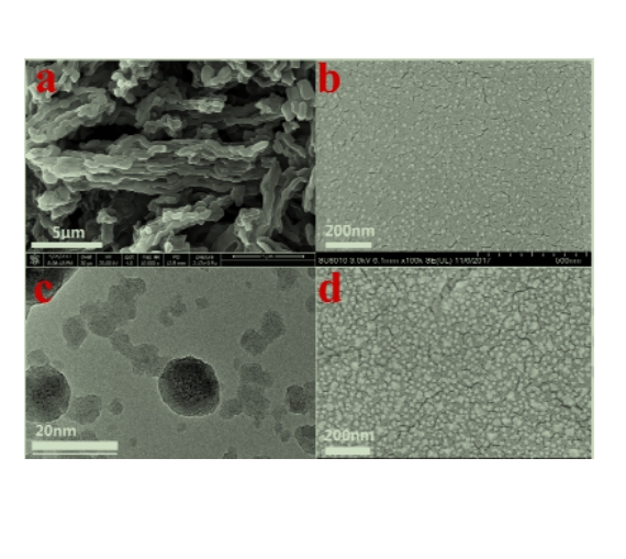
图为:(a)OMC-CS的扫描EM图像;(b)SA-AuNPs的扫描EM图像;(c) SA-AuNPs+Bio-DNA1的TEM图像;(d)SA-AuNPs+Bio-DNA1的扫描EM图像。
结论:由于AuNPs-SA复合材料具有良好的电导率,在加入AuNPs-SA复合材料后,AuNPs-SA/OMC-CS/GCE复合材料的峰值电流表现良好。OMC-CS 193和AuNPs-SA复合材料为该适触点传感器提供了电子传递能力。当Bio-DNA1被固定在AuNPs-SA/OMC-CS/GCE表面时,由于寡核苷酸的非导电性,峰值电流降低。